Daijiworld Media Network - New Delhi
New Delhi, Oct 25: Combining subtraction iodine maps (SIM) with deep learning reconstruction (DLR) can improve computed tomography (CT) image quality and visualisation of bone involvement in patients with temporal bone lesions, according to a prospective study.
The technique outperformed conventional contrast-enhanced CT using hybrid iterative reconstruction (HIR), with particularly significant improvements in lesions showing high contrast enhancement.
Researchers evaluated 77 patients with temporal bone lesions who underwent contrast-enhanced CT scans. They compared four imaging techniques: conventional enhanced CT reconstructed using HIR, enhanced CT using DLR, SIM combined with HIR, and SIM combined with DLR.
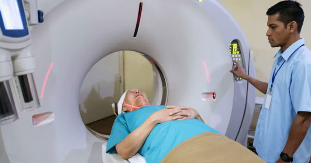
SIM images were generated by subtracting pre-contrast images from contrast-enhanced images and overlaying the resulting maps onto bone-window images, allowing researchers to assess the lesions more clearly.
Two radiologists independently evaluated overall image quality, confidence in detecting lesions, delineation of lesion contours and visualisation of temporal bone involvement. Quantitative measurements, including image noise and contrast-to-noise ratio (CNR), were also compared.
The findings showed that SIM combined with DLR significantly improved all four qualitative assessment measures compared with conventional contrast-enhanced CT using HIR. The combined technique also reduced image noise and increased the contrast-to-noise ratio of lesions.
The researchers reported strong agreement between the two radiologists. The benefits were particularly evident in lesions with high contrast enhancement, which showed greater improvements in detection confidence, contour delineation and visualisation of bone involvement than lesions with low enhancement.
The researchers concluded that combining SIM with DLR could improve image quality and anatomical delineation, helping clinicians better assess the extent of temporal bone lesions before surgery and plan appropriate treatment, particularly in highly vascular lesions.
However, the study had limitations, including its single-centre design and relatively small sample size. The researchers also noted that the performance of deep learning reconstruction algorithms may vary between CT manufacturers, potentially limiting the wider applicability of the findings.
Larger, multicentre prospective studies are needed to validate the clinical reliability of the technique and establish its broader role in assessing temporal bone lesions.