Daijiworld Media Network – Los Angeles
Los Angeles, Jan 16: In a major breakthrough for medical diagnostics, researchers from the Keck School of Medicine of the University of Southern California (USC) and the California Institute of Technology (Caltech) have demonstrated a novel, noninvasive imaging technique capable of capturing fast, detailed 3D images of the human body from head to toe.
The proof-of-concept study, funded by the US National Institutes of Health and published in Nature Biomedical Engineering, combines ultrasound with photoacoustic imaging — a technology that detects sound waves produced when light interacts with blood. The hybrid approach allows simultaneous visualisation of body tissues and blood vessels within seconds.
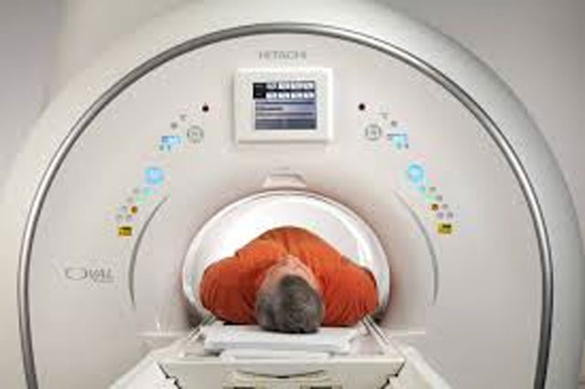
Medical imaging plays a vital role in diagnosing injuries, infections, cancer and chronic diseases. However, existing tools such as ultrasound, X-rays, CT scans and MRI often face limitations related to cost, scan time, radiation exposure, image depth and field of view.
“You cannot understate the importance of medical imaging for clinical practice. Our team identified critical limitations of current techniques and developed a novel approach to address them,” said Dr Charles Liu, professor at the Keck School of Medicine and co-senior author of the study.
To demonstrate the versatility of the new system, researchers successfully imaged multiple body regions, including the brain, breast, hand and foot. Brain scans were conducted on patients undergoing surgery for traumatic brain injury, where part of the skull had been temporarily removed. The system was able to capture tissue structures and blood vessels across areas up to 10 centimetres wide in just about 10 seconds.
The new platform, named RUS-PAT (Rotational Ultrasound and Photoacoustic Tomography), integrates two advanced imaging methods. Rotational ultrasound tomography uses an arc of detectors to generate 3D images of tissues, while photoacoustic tomography employs laser light absorbed by blood to produce sound waves that map blood vessels in three dimensions.
“This method allows far more comprehensive imaging at meaningful depths. It is an exciting step forward in noninvasive diagnostics without the use of ionising radiation or strong magnets,” said Dr Lihong Wang of Caltech, co-senior author of the study.
Compared to MRI and CT scans, RUS-PAT is less expensive to build, avoids radiation exposure and delivers more detailed images than conventional ultrasound.
By successfully imaging different parts of the body, the researchers highlighted the technology’s wide clinical potential — from diagnosing stroke and neurological disorders to improving breast cancer detection and managing diabetic foot and vascular diseases.
Despite the promising results, researchers cautioned that further refinement is needed before the technology can be used in routine clinical practice. One key challenge remains imaging through the intact human skull, which can distort signals. Ongoing work aims to overcome this by adjusting ultrasound frequencies and improving signal processing.
“This is an early but important step. We are continuing to refine the system as we move closer to future clinical use,” Dr Liu added.