Daijiworld Media Network - New Delhi
New Delhi, Aug 6: Exercise stress echocardiography (ESE) may help identify people at risk of developing acute mountain sickness (AMS) within hours of arriving at high altitude, according to a new study.
Researchers found that specific changes in right ventricular function and pulmonary circulation during exercise accurately distinguished participants who later developed acute mountain sickness from those who remained symptom-free.
Acute mountain sickness commonly affects people travelling to elevations above 2,500 metres and can cause symptoms such as headache, nausea, dizziness, fatigue and sleep disturbances. Although several risk factors have been identified, clinicians currently have limited methods to predict who will develop the condition before symptoms appear.
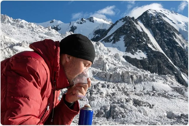
The prospective study involved 50 healthy adults living at low altitude who travelled by bus to an elevation of 3,600 metres.
Within six hours of arrival, participants underwent exercise stress echocardiography to assess right ventricular function and pulmonary haemodynamics. They were evaluated for acute mountain sickness the following morning using the Lake Louise Score.
Of the 50 participants, 23 (46 per cent) developed acute mountain sickness, while the remaining 27 showed no symptoms.
The study found that participants who later developed acute mountain sickness had significantly higher systolic pulmonary artery pressure (SPAP) and pulmonary vascular resistance (PVR) during peak exercise than those who remained symptom-free.
They also experienced greater increases in SPAP and PVR from resting levels during exercise, indicating a stronger pulmonary vascular response to high altitude.
In contrast, measures of right ventricular contractile reserve, including tricuspid annular peak systolic velocity (TV s′) and right ventricular fractional area change (FAC), showed smaller improvements during exercise among participants who developed AMS, suggesting reduced right ventricular functional reserve.
Researchers also observed larger inferior vena cava (IVC) diameters in the AMS group at both rest and peak exercise.
Based on the physiological measurements obtained during exercise testing, the researchers developed a predictive model incorporating peak IVC diameter, changes in pulmonary vascular resistance (ΔPVR) and changes in TV s′.
The model demonstrated strong predictive performance, achieving an area under the receiver operating characteristic curve (AUC) of 0.865 and correctly identifying participants with an overall accuracy of 84 per cent.
According to the researchers, combining measurements of pulmonary vascular response and right ventricular function could provide a practical approach for identifying individuals at increased risk of developing acute mountain sickness shortly after reaching high altitude.
The authors said exercise stress echocardiography can detect early cardiovascular changes associated with acute mountain sickness even before clinical symptoms become apparent.
They added that early identification of abnormal pulmonary vascular and right ventricular responses could help clinicians implement preventive measures, improve monitoring and guide decisions on whether individuals should continue ascending to higher altitudes.
The researchers emphasised that larger studies are required to validate the predictive model and determine how exercise stress echocardiography can be incorporated into routine assessments for travellers, athletes and workers operating at high altitudes.