Daijiworld Media Network - New Delhi
New Delhi, Apr 17: Contrast-enhanced mammography (CEM) demonstrates performance comparable to MRI in assessing tumour size and detecting additional malignant lesions, with closer agreement to histopathology in evaluating disease extent, according to a retrospective analysis.
CEM combines conventional mammography with contrast enhancement to improve visualisation of tumour vascularity. In breast cancer imaging, accurate estimation of tumour size and spread is crucial for surgical planning and identifying multifocal or multicentric disease.
The study, conducted on 52 women with biopsy-confirmed breast cancer, found high agreement between CEM, MRI and histopathology. Two radiologists independently reviewed imaging studies while blinded to pathology results, minimising bias.
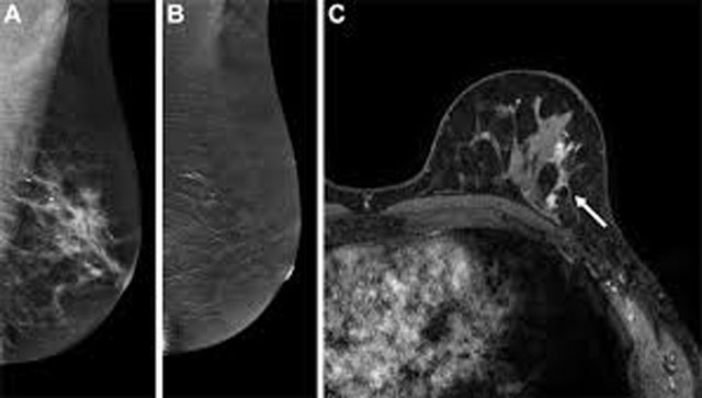
CEM detected 51 of the 52 primary tumours, while MRI identified all cases. Tumour size measurements were nearly identical, with an average of 24.9 mm on CEM and 25.2 mm on MRI, indicating strong correlation between the two methods.
When compared with post-operative histopathology, CEM showed exact agreement in assessing overall disease extent, both averaging 32.6 mm. MRI, however, slightly overestimated the extent, with a mean measurement of 35.0 mm. Larger discrepancies were observed in a few cases, particularly those involving non-mass enhancement, which can be more challenging to interpret.
The findings suggest that CEM could serve as a viable alternative to MRI for preoperative evaluation in selected patients. However, experts caution that its limitations must be recognised, as even a small number of missed lesions can significantly impact treatment planning.
While MRI remains more sensitive in detecting all lesions, the study highlights the growing potential of CEM as an efficient and accessible imaging option in breast cancer care.